Toolkits menu: 
The Cord Finder tool can be used to isolate the spinal cord on MRI spinal cord scans, and to calculate the cross-sectional areas along the cord, perpendicular to the cord axis. In addition, there is a specialised tool ( Cord Follow-Up) for estimating the change in atrophy between two time-points.
Start the Cord Finder tool from the Toolkits menu: 
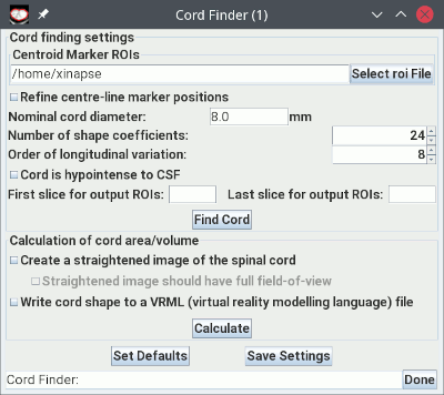
The Cord Finder tool will now appear as shown below:

Note that the ROI Toolkit also pops up when you start the Cord Finder.
The procedure for outlining the cord and calculating the cord areas can be summarised as follows: